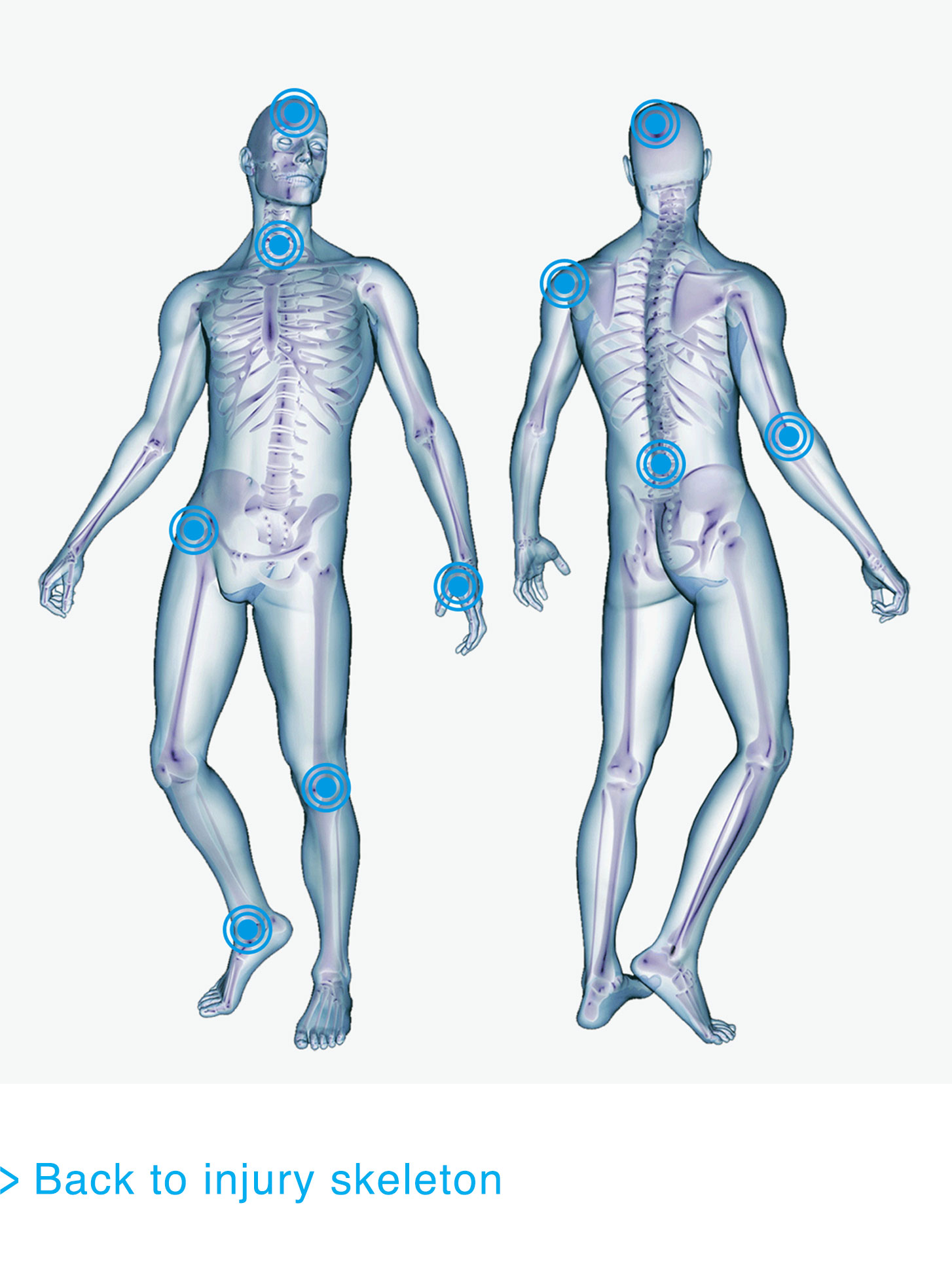
Looking after your knees is crucial; knees are the largest joint in the human body – and also the most likely to get injured.
Fascinating facts
- A common myth is that regular running increases your chance of getting osteoarthritis in your knees, but there is no evidence to support this.
- To look after your knees, keep the weight off. Every extra pound we carry places uneccessary extra strain on them.

Related injuries
The Anterior Cruciate Ligament (ACL) is one of four ligaments in the knee. It runs from the back edge of the bottom of the thigh bone (femur) to the front edge of the top of the shin bone (tibia).
The job of the ACL is to provide stability to the knee by stopping the tibia from sliding forward or twisting.
The symptoms of an ACL tear are the immediate onset of pain, a snapping noise, immediate swelling, loss of function and stability in the knee.
ACL injuries usually occur when the knee twists very quickly, the person lands awkwardly or suddenly slows down with the foot planted, placing excessive forces on the ACL causing it to tear. The ACL is the most commonly injured knee ligament and every year in the UK there are 30 ACL injuries for every 100,000 people.
Occurrence/Frequency of Injury
ACL tears commonly occur during sporting activities such as football, rugby, and basketball due to the dynamic nature of these sports, and are only caused by contact (i.e. a tackle) in 20% of cases. A tear is more likely to occur during a turning or pivoting movement. The ACL tear is caused when the ligament can no longer cope with the force generated by the twisting or forward movement of the knee.
Orthopaedic consultants and physiotherapists can use a variety of tests to indicate that an ACL has been injured, such as the anterior draw test, but the only way to truly confirm the injury is by using an MRI scan.
ACL tears don't occur in isolation and can be associated with meniscal tears and collateral ligament damage, making recovery even more challenging.
Treatment and Recovery Timeframes
Those who have suffered a partial tear to the ACL shouldn't experience any lasting issues and will be able to return to normal activity within eight weeks. However, exercises to strengthen the knee are strongly recommended to help prevent the chance of a full rupture occurring.
If a full rupture has occurred and it is deemed that greater knee stability is required, for example to support a return to sport, an ACL reconstruction may be required. The ligament is reconstructed using another tendon or ligament to substitute for the torn ligament.
Effective rehabilitation following the injury is crucial to ensure a successful outcome as the replacement tendon cannot provide the same stability and on average recovery takes at least nine months.
Rehabilitation following ACL surgery focuses on restoring motion and strength, and improving the stability of the joint to prevent future injuries. Therefore, following the reconstruction, it is crucial that suitable physiotherapy is put in place because, if it isn't, the reconstruction may fail.
Interesting Facts
Sorry ladies! Evidence shows that women are five times more likely to suffer an ACL tear than men.
Anterior Knee Pain is an umbrella term which encompasses a wide range of related but significantly different conditions resulting in pain around or behind the knee cap. 25% of the population will be affected by it at some time and it is the most common over-use syndrome affecting sports people - although you do not have to be particular sporty to suffer from the injury. The two most common causes are patella-femoral syndrome and quadriceps tendonitis.
Occurrence/Frequency of Injury
Patella-femoral Syndrome occurs when the under-surface of the patella (knee cap) becomes pain sensitive, due to damage and irritation to any of the structures between the patella and femur. This can be caused by incorrect tracking or movement of the patella as the knee bends and straightens. The patella rubs against the femur instead of gliding correctly in the groove.
Patella Tendonitis refers to a painful over-use injury of the patella tendon, which connects the knee cap (patella) to the shin (tibia). This occurs as a result of degeneration (either acute or chronic) and a 'weakening' within the patella tendon itself, without the presence of inflammation. Many cases of patella tendonitis also occur in conjunction with patella femoral syndrome.
Treatment and Recovery Timeframes
The timeframe for recovery is very much dependent on the level of damage or irritation. The first step in the recovery process is to rest and give sufficient time for the inflammation to settle down. During this period regular ice and pain killers will help manage the associated symptoms.
Following this, a quadriceps strengthening program is needed to increase the strength of the patella tendon. This involves a graduated series of exercises, that start slowly, and then progress by increasing the speed and weight involved during exercise. A stretching programme is also essential because poor flexibility can overload the hamstring and quadriceps.
Anterior Knee Pain is unlikely to be caused by work activities unless it involves long periods of kneeling or strenuous activities. Therefore, the period of absence from this type of injury should be minimal. However, it is important that modified duties ways of working are considered for people suffering from this type of injury.
Interesting Facts
Many people believe that running increases your chances of suffering from osteoarthritis in later life, but this is in fact false. Many studies have looked at this and no link has been found between running and the development of osteoarthritis so dust off your running shoes and get out there........just don't forget to warm up your knees!
There are many different types of cartilage in the human body - one type (Hyaline) covers the ends of our bones and its job is to form a smooth congruent surface so the joint surfaces move smoothly over each other.
Cartilage Tears can occur in any of our joints where it is present, but tears are most common in our knee. The cartilage in the knee is called the meniscus and works as a shock absorber to protect our knee.
Occurrence/Frequency of Injury
Cartilage tends to be damaged by impact and or twisting injuries. Every year in the UK, around 10,000 people have cartilage damage serious enough to require treatment.
Cases of accidental cartilage damage are most common in people under 35 years old. This is because this age group is more likely to take part in sporting activities, which greatly increase your risk of suffering this type of injury.
Treatment and Recovery Timeframes
Both meniscus and hyaline (also know as chondral cartilage) can be damaged. Hyaline cartilage does have some potential to heal itself if it has a good blood supply. Meniscus however generally does not heal as it is sandwiched between two bones and the blood supply is poor. So surgery is often required if conservative treatments fails.
If surgery is advocated, it is done by keyhole surgery (otherwise known as an arthroscopy). The surgeon will remove the damage and repair any frayed edges of the cartilage.
Physiotherapy can begin quickly after surgery. The main focus is to decrease swelling, regain full range of motion and build muscle strength.
Rehabilitation varies greatly depending on the type of injury, but the typical rehabilitation programme includes a short period in a knee brace, performing non weight-bearing and range of motion exercises. After this, patients can begin weight bearing and other exercises. Over time, low impact exercise can begin with a final progression on to full activity as and when the person is ready to do so.
Interesting Facts
Menicus surgery often involves removing or repairing the damaged cartilage. Damage to hyaline cartilage on the bone's surface, if symptomatic, is often treated by a surgical procedure know as micro-fracture. In this case, the bone is drilled to cause bleeding and hopefully stimulate the healing and re-growth of cartilage.
A ligament is a short band of tough, flexible tissue, made up of lots of individual fibres, which connect the bones of the body together. Collateral ligaments are found inside and outside of many of our joints including our elbows, fingers and thumbs.
The knee joint has one on either side with the one on the inside of the joint being known as the medial collateral ligament and the one on the outside, called the lateral collateral ligament.
Occurrence/Frequency of Injury
A collateral ligament can be partially torn but may still remain fairly stable. This is because a collateral ligament is not a single, thin ligament, but consists of many minor ligaments in different directions and layers, which mesh together into one single strong ligament. Injuries to the collateral ligaments are usually caused by a force that pushes the knee sideways and commonly occur as a result of contact injuries. Sufferers will complain of pain on the outside of the knee and there is usually evidence of swelling.
A grading scale is used to convey the severity of a ligament injury. The degree of the symptoms felt, often correlating with the extent of the damage sustained:
Grade I Sprain: The symptoms tend to be limited to pain and swelling. Most people can walk without crutches, but may not be able to jog or jump.
Grade II Sprain: There is usually more significant swelling and bruising caused by bleeding under the skin. People often have pain with walking, but can take a few steps.
Grade III Sprain: The joint is usually quite painful, and walking can be difficult. People may complain of instability, or a giving-way sensation in the joint.
Treatment and Recovery Timeframes
For a Grade I sprain, use RICE (rest, ice, compression and elevation):
- Rest and avoid putting strain through the joint.
- Ice should be immediately applied as it keeps swelling down. It can be used for 20 minutes to 30 minutes, three or four times daily.
- Compression dressings or bandages immobilise and support the injured area.
- Elevate, when able, above your heart level for 48 hours.
For a Grade II sprain, the RICE guidelines can also be used. It will take longer to heal and the patient may be provided with a brace to immobilise the joint.
A Grade III sprain can lead to some level of instability and surgery may be required, particularly if the ligament has completely ruptured.
There are usually three phases of recovery, which should be followed to achieve maximum recovery:
- Phase 1 includes resting, protecting the joint and reducing the swelling (one week).
- Phase 2 includes restoring range of motion, strength and flexibility (one to two weeks).
- Phase 3 includes gradually returning to activities that do not require turning or twisting of the joint and progressing maintenance exercises (weeks to months).
Grade I and mild Grade II sprains should not require much time off work (on average no more than one week), but more severe sprains may require a longer period off to allow the ligament to repair, which is often supported by a period of immobilisation.
Interesting Facts
Ligaments take a lot longer to heal than muscles due to their poorer blood supply.
Hamstring injuries are a result of damage to the fibres of one of the three muscles that make up the hamstring- biceps femoris, semitendonosus and semimembranosus. Hamstring injuries are classed from a Grade I tear (damage to a few muscle fibres) to a Grade III tear - or a full rupture of all or some of the muscles mentioned above.
Occurrence/Frequency of Injury
Hamstring injuries are usually associated with sports such as rugby and football as they involve lots of running and occasional sprinting. This is because the two most common causes - fatigue and muscle imbalance- arise from these sorts of activities. The hamstring has to work hard during running and sprinting to control the power generated by our quadriceps. When the hamstring can no longer do this, the muscle will tear at a tension point.
Hamstring injuries are less likely to occur at work as most jobs don`t involve sprinting or running. However, they can occur when an individual falls or trips as the muscle is stretched beyond its elastic limit.
Treatment and Recovery Timeframes
Initially hamstring tears needs to be treated with RICE (rest, ice, compression and elevation). A Grade I tear won`t require excessive treatment, just a period of rest from exercise. Recovery would be expected in 2-10 days, with time off work not required.
In more severe cases (those with a grade II strain), the length of time off sport will increase (10 days-6 weeks) and the initial goal, once swelling and pain has decreased, is to restore the full range of movement and gradually increase strength. It is important that the hamstring is strengthened to prevent recurrence, especially if the injury was caused by sporting activity. Treatment for more severe strains remains the same but recovery takes longer. When a full rupture has occurred a period on crutches to enable the fibres to heal or surgery may be necessary. In these cases recovery timeframes will be extended.
Only those with Grade III or severe Grade II tears require time off work. Even in these cases an employee would be expected to return within 28-56 days.
Those returning to sport need to ensure they have allowed sufficient time for the fibres to heal and the muscle to strengthen. Injury re-occurrence is often as a result of returning to sport too soon after injury, causing further damage and longer absence from normal activity, which can be very frustrating.
Interesting Facts
In a recent survey by the RFU, hamstring tears were found to be the second most common injury among rugby players. If you regularly play sport you need to look after your hamstrings!
Get in touch
If you want to know more about our services or you have a rehabilitation issue to discuss, please get in touch via postclaimrehab@uk.qbe.com