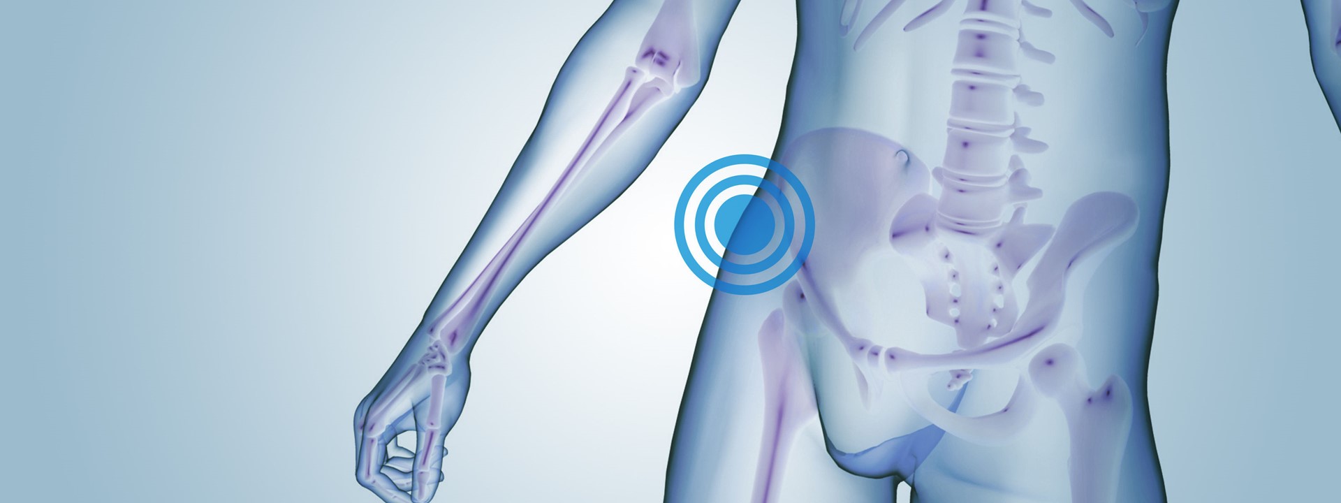
The primary function of the hip joint is to support the weight of the body in static and moving postures. Together, the hip joints are the most important part of the body for retaining balance.
Fascinating facts
- The hip is the second most flexible joint in the body (behind the shoulder).
- Like a giant shock absorber, each hip joint must be able to manage half of the body's weight.
- During running and jumping, the force of the body's movement multiplies the force on the hip to many times the normal force applied by the body.
Related injuries
Hip Bursitis (also called trochanteric bursitis) causes pain on the side of the hip making it uncomfortable to lie on the affected side.
Quite simply, bursitis is the inflammation of the bursa which is a fluid filled sac that protects the soft tissues of the joint. When the hip bursa becomes inflamed, it causes pain in the hip which can radiate down towards the knee.
Bursa are normally found around bony points such as the hip, knee and the elbow. The trochanteric bursa is located on the lateral side of the leg over the bony prominence of the hip.
Occurrence/Frequency of Injury
There is some evidence that women are four-times more likely to suffer from hip bursistis than men.
The condition can be diagnosed by a physiotherapist through manual tests and also ultrasound imaging.
Causes for bursitis can range from muscle imbalance, trauma or associated joint wear and tear.
Treatment and Recovery Timeframes
Treatment for Hip Bursitis aims to settle the inflammation in the first instance. It is managed by physiotherapy treatment including ice, electrotherapy and stretching exercises.
If the bursitis has been caused by an accident such as a fall, the inflammation can resolve relatively quickly within 1-2 weeks. Only those where there is an underlying cause does it take longer to resolve, as the primary problem has to be addressed first before the secondary bursitis pain can resolve. In chronic conditions, a corticosteroid may be necessary to settle the inflammation.
Athletes often develop bursitis owing to tight structures repeatedly rubbing over the hip's bony prominence and irritating the bursa. Treatment aims to address these muscle and tissue imbalances by stretching, muscle retraining and often a period of rest or reduced activities.
Some cases of bursitis do not resolve with conservative treatment and require surgical intervention - but these cases are in the minority.
Interesting Facts
If the condition continues, the inflammation can become chronic and the bursa is at risk of calcifying (turning to bone) and at this point surgery is the only option- therefore early treatment and medical advice is recommended.
A Hip Dislocation occurs when the head of the femur slips out of its socket in the hip bone (pelvis). In approximately 90% of incidents, the femur is pushed out of its socket in a backwards direction (posterior dislocation). An anterior dislocation occurs when the femur slips out of the socket in the forwards direction.
Hip Dislocation is very painful and patients will be unable to move the leg and, if there is nerve damage, may not have any feeling in the foot or ankle area.
Occurrence/Frequency of Injury
Dislocations are caused when a significant force is applied to the joint causing the head of the femur to come out of the hip socket.
One of the most common causes seen by the insurance claims industry is road traffic accidents when the abdomen is suddenly moved towards the knees causing an extreme force to be applied to the hip. Other causes include falls from ladders or generally from height.
Treatment and Recovery Timeframes
Due to the risk of nerve damage and reduced blood supply to the leg, medical assistance should be sought immediately. Treatment will involve administering a sedative or geneal anaesthetic to manipulate the head of femur into the socket by either sedative or general anaesthetic in surgery and should occur as soon as possible.
Once the surgeon is happy that the femur is correctly aligned, physiotherapy treatment can take place to work on returning to normal walking pattern, mobility and strength to the hip joint. Physiotherapy and a suitable exercise programme are essential to achieve a full recovery.
If an uncomplicated dislocation has occurred, recovery will usually take place within three months. A return to work to heavy manual positions should be guided by the treating team. However, once the hip is strong enough and the risk of further dislocation is reduced, a return to work can occur.
Complications may include nerve and vascular damage to the lower limb which will require additional treatment which will extend the recovery and return to work.
Interesting Facts
Only 5% of Hip Dislocations occur through sporting activity.
A Hip Fracture is a 'break' to the joint between the femur and the pelvis, which is a ball and socket joint. The ball (head) of the femur fits into the socket (known as the acetabulum) of the pelvic bone to make the hip joint.
There is a strong but flexible joint capsule that surrounds the hip joint. It helps to give stability and produce synovial fluid, which provides lubrication that helps with joint movement.
Occurrence/Frequency of Injury
There are 75,000 reported Hip Fractures in the UK each year with 80% of these involving elderly women who are at risk of osteoporosis.
The other 20% of injuries comes from falls from height and direct trauma (for example, a road traffic accident).
Treatment and Recovery Timeframes
The fracture is normally diagnosed by an Orthopaedic Consultant through a review of X-Ray investigations. Depending on the severity, a decision will be made on appropriate management.
Most Hip Fractures require orthopaedic surgery to insert an orthosis to stabilise the fracture and allow healing of the bone. This can range from the surgeon inserting screws and plates through the fracture site, to replacement of the head of femur with a prosthetic implant (hip replacement).
Following surgery, and depending on post-surgical precautions (bed rest may be indicated initially), effective rehabilitation is key to recovery. Physiotherapy will usually take place the day after surgery to begin to mobilise with walking aids. This is also important to prevent complications related to prolonged bed rest such as pressure sores and deep vein thrombosis. A patient may also be seen by an occupational therapist to promote independence in activities of daily living.
Physiotherapy sessions will be more intensive post surgery whilst in hospital and will reduce over a three month period to the injured party being able to self-manage their rehabilitation. A focused gym and swimming pool based personal training programme may assist in recovery as return to work becomes a focus.
The return to work is guided by the Orthopaedic Surgeon or GP. Usually, healing of the bone is complete at six months when a person will be deemed fit to work. However, if the patient is able to return to work on sedentary duties earlier, then this should be discussed at the earliest opportunity.
Interesting Facts
In January 2010, at the age of 62, the dancer Wayne Sleep underwent hip replacement surgery. Three months later he was dancing the role of one of the Ugly Sisters with the Royal Ballet. Part of his routine consisted of 50 continuous jumps - in high heels! Impressive stuff!
A labrum is found in both the shoulder and the hip joint and forms a cartilage-like-ring around the edge of the bony socket of the joint. It helps to provide stability to the joint by deepening the socket, yet unlike bone, it also allows flexibility and motion. Normally the surface of the labrum is very smooth, allowing easy movement of the femur on the pelvis. Occasionally the labrum can be torn or damaged so that the surface is no longer smooth. When this occurs, the condition is known as a labral tear.
Occurrence/Frequency of Injury
A labral tear often occurs traumatically when impact causes the leg to change direction suddenly (for example when falling from height or when involved in a car accident). Occasionally, the condition may develop over time through gradual wear and tear associated with excessive twisting or weight bearing activity. In older people, degenerative changes to the hip joint may also be present. In these instances, injury to the labrum may occur with a relatively trivial movement. Injury is characterised by a deep throbbing pain affecting mobility and sleep and also reduced range of movement. A clicking sensation in the hip joint can often be present.
Treatment and Recovery Timeframes
This injury rarely occurs in isolation and so there is often damage to other parts of the hip structure. Firstly, to rule out any bony injury, an X-ray is required. If this shows no bony injury, then an MRI will be performed to look at the soft tissue structures. Non-surgical treatment can include any of the following which are used to alleviate pain and to maximise range of movement to allow the injury to settle conservatively:
- Anaesthetic injection into the hip joint
- Anaesthetic medication
- Physiotherapy to increase hip strength, mobility and stability.
Most people recover with the above but, if hip pain is experienced for more than 12 weeks with physiotherapy treatment, surgery may be required. In some cases surgery may be recommended immediately if symptoms of hip locking are present. In most cases the surgery will be an arthroscopic procedure during which the surgeon will remove the torn tissue or elect to sew it back together. Following surgery, recovery can take between 6 to 12 weeks depending on the nature of the tear. During recovery, medical precautions to avoid high impact and strenuous activities are likely to be placed on the patient. Some sessions of physiotherapy will be required (usually no more than eight) which may be aided by a focused gym programme. In most cases, a full return to function is likely. However, if the labral tear is affected by degenerative changes (ie in older people), a return to heavy manual work is not recommended given the risk of re-injury.
Interesting Facts
More prevalent in older people as their hip stability reduces with the degeneration process.
Warming up correctly prior to strenuous exercise may help prevent damage if sudden changes of direction of the hip joint are likely to be encountered.
If you run, cycle or are pregnant, you may or you may not want to read about Iliotibial Band Syndrome ( ITBS).
ITBS is the irritation of the Illiotibial band (ITB) where it joins the knee, resulting in tension and tightness. Chances are, if you are a runner, you have encountered this injury before or at least know of people who have.
Occurrence/Frequency of Injury
The ITB is a superficial thickening of tissue on the outside of the knee that extends from the outside of the pelvis, over the hip and knee, and attaches just below the knee. It is not a strong structure so if the surrounding muscles are weak you are at risk of developing ITB syndrome. Running requires a stable knee, we often do not focus on specific exercises that ensure the knee is strong, leading to anatomical abnormalities or muscular imbalances. The instability leads to minor injuries and tightness along the lateral (side of) leg and the development of ITBS. Pregnant women may also suffer ITBS where the band connects to the hip - hormonal changes result in the tissue becoming looser, ready for birth.
Treatment and Recovery Timeframes
With acute pain, the ITB can be rested, iced, compressed and elevated (RICE for short).
Stretching, stretching, stretching - use of foam rollers can help. A patient will need to continue to stretch even after the pain has gone as it has a nasty habit of coming back.
Physiotherapy in the form of ultrasound and taping can help, but effort should also be paid to identify the underlying cause and correct it e.g. muscle imbalance.
If pain fails to resolve, pain killer injections can be administered to settle the joint down.
The injury should not lead to any time off work. Only those involved in jobs that require a lot of walking and climbing stairs may require a short period of modified duties to allow the symptoms to settle.
Interesting Facts
If you have ITBS you'll need, at least during acute phase, to avoid running, mountaineering, dancing, weight lifting, rugby, rowing, football, cycling, gymnastics, bowling... the list goes on, otherwise the injury will continue to get worse.
A top tip if you're a runner is to replace your running shoes regularly. A well used running shoe will not provide the support you require and will result in knock-on consequences further up your leg, the development of ITB syndrome being one of them.
Get in touch
If you want to know more about our services or you have a rehabilitation issue to discuss, please get in touch via postclaimrehab@uk.qbe.com